Technological Advancements in Brain Tumor Surgery

Brain tumors are the toughest and demanding forms of cancer to cure due to their presence on the brain and sensitive nature of brain tissue. Moreover, technological evolution in brain tumor surgery has evolved the field, giving new scope for patients and enhancing results. This article ranges over the new developments in surgical techniques, picture technologies, and cure procedures that are changing brain tumor surgery.

Introduction

The stream of brain tumor surgery has seen good evolution over the past few years, catalyzed by a good research of brain tumor biology and the advancement of new technologies. These improvements have evolved the precision of surgical innovations, increased the potential of treatments, and lowered the risk involved with brain surgery. This article involves the latest technological research that is crafting the future of brain tumor surgery, providing the new hope for patients and healthcare professionals.

Enhanced Imaging Techniques

Among the most critical characteristics of brain tumor surgery is the potential way to locate and analyze the tumor. Improvements in neuroimaging have played a pioneering role in this sector. Magnetic resonance imaging (MRI) and positron emission tomography (PET) have become important tools for neurosurgeons. MRI provides complete anatomical information, while PET provides overview into the metabolic activity of brain tumors. Simultaneously, these imaging methods allow for exact mapping of the tumor and surrounding brain structures, providing safer and potential surgical innovations.

An important innovation in this stream is the involvement of multimodal imaging techniques.Involving MRI with newest molecular imaging, such as PET, improving the capacity to differentiate between tumor tissue and healthy brain tissue. This is specifically considered for planning surgical resections and lowering harm to critical brain sectors.

Yearwise Publication Trend on neuroimaging

Find publication trends on relevant topics

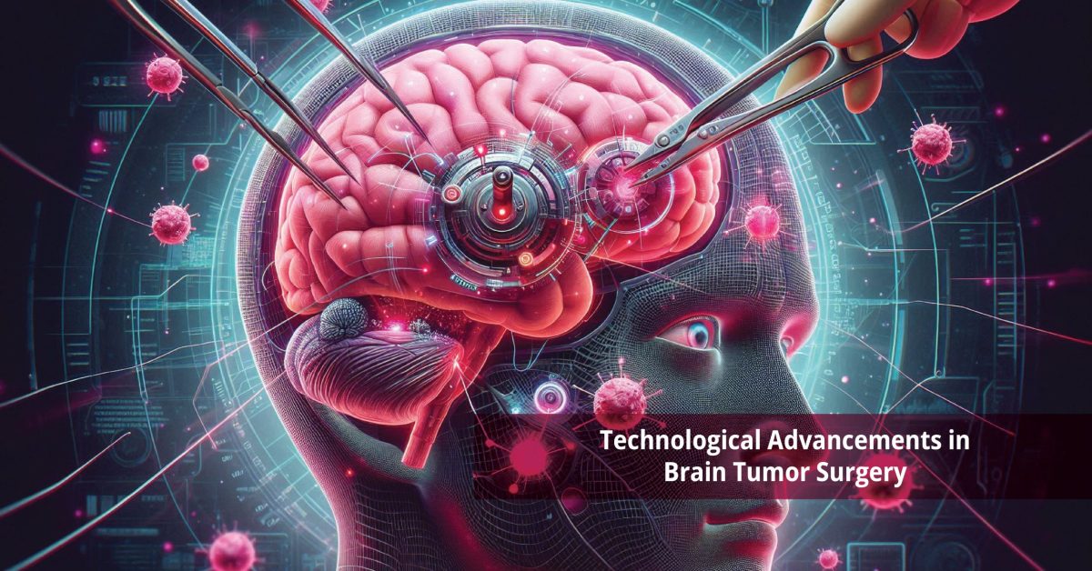
Intraoperative Imaging

Intraoperative imaging has evolved the accuracy and safety of brain tumor surgeries.Technologies like intraoperative MRI (IMRI) and intraoperative ultrasound (iUS) have real – time imaging during surgery, permitting surgeons to analyze the tumor and surrounding brain structures as they operate. This real – time input helps confirm complete tumor removal while conserving as much healthy brain tissue as possible.

IMRI, for instance, has been shown to enhance the extent of tumor resection and decrease the likelihood of leaving behind residual tumor tissue. This technology allows surgeons to make immediate adjustments during surgery, scoping for good outcomes for patients.

Image-Guided Surgery

Image – guided surgery (IGS) systems have become important tools in the recent era of brain tumor surgery. This mechanism makes use of preoperative and intraoperative picture scanning data to create a 3D map of the patient’s brain. Surgeons can then use this map to route through the tumor, keeping away critical brain structures and leading surgical accuracy.

Most important components of IGS are neuronavigation, which gives real- time inputs on the surgeon’s tools location within the brain. This technology increases the surgeon’s potential to evict the tumor while lowering damage to surrounding healthy tissue. Moreover, the involvement of augmented reality (AR) into the IGS mechanism is an upcoming trend, providing surgeons increased visualization and spatial awareness during the process.

Minimally Invasive Techniques

Minimally invasive surgical innovations have transfigured brain tumor surgery by decreasing the invasiveness of process, transforming to more efficient recovery times and lesser complications. Techniques such as endoscopic surgery and laser consumption have achieved popularity in recent years.

Endoscopic surgery involves the use of a small camera and specialized tools inserted through a tiny incision, permitting surgeons to enter and eliminate tumors with lesser disruption to surrounding brain tissue. This process is mainly beneficial for tumors located in hard – to – reach areas.

Laser ablation, on the other hand, uses laser energy to completely target and remove tumor cells. This technique is especially useful for curing small, deep – located tumors that are hard to access with traditional surgical processes. Laser depletion offers the benefit of being less invasive and involved with shorter hospital stays and fast recovery period.

Advanced Surgical Instruments

The development of new surgical tools has further increased the precision and performance of brain tumor surgeries. Robotic – assisted surgery, for example, provides for bigger control and additionally, the utility of fluorescence – mediated surgery has become more common. This procedure involves the injection of fluorescence – guided surgery has become more common. This technique involves the injection of a fluorescent dye that piles up in tumor tissue, making it glow under specific lighting conditions. Surgeons can then visualize the tumor more clearly and differentiate it from healthy brain tissue, confirming more complete tumor removal.

Targeted Therapies and Personalized Medicine

The invention of targeted therapies and customized medicine has brought about a paradigm transfer in the cure of brain tumors. Traditional chemotherapy and radiation therapy often have limited efficiency against brain tumors and can cause significant side effects. Moreover, targeted cures that specifically target tumor cells while sparing healthy tissue offer a more efficient and less toxic alternative.

One important example is the use of bevacizumab, an anti-angiogenic drug that restricts the formation of new blood vessels in tumors, efficiently starving them of nutrients and oxygen. Clinical trials have shown that bevacizumab, either alone or in combination with other drugs, can enhance survival rates in patients with recurrent glioblastoma, an especially aggressive type of brain tumor.

Customized medicine, which involves treatment based on the genetic profile of the tumor, is also adding traction. By recognizing specific genetic mutations and molecular markers in a patient’s tumor, oncologists can opt for the most suitable targeted therapies, enhancing treatment results and decreasing side effects.

Recent Publications on neuroimaging

Find publications on relevant topics

Immunotherapy

Immunotherapy is another prompt area of research in brain tumor treatment. This process harnesses the body’s immune system to identify and target cancer cells. Different immunotherapy techniques, involving immune checkpoints inhibitors, CAR-T cell therapy, and cancer vaccines, are being developed for their effective treatment of brain tumors.

Clinical trials have demonstrated the potential of immunotherapy in improving outcomes for patients with brain tumors. For instance, immune checkpoint inhibitors, which block proteins that inhibit the immune response, have shown promise in treating certain types of brain tumors by enhancing the body’s ability to target and destroy cancer cells.

Conclusion

Technological improvements in brain tumor surgery have transformed the landscape of treatment, offering new scope to patients and improving outcomes. More effective  imaging techniques, intraoperative imaging, image-guided surgery, minimally invasive techniques, innovative  surgical instruments, targeted therapies, personalized medicine, and immunotherapy are all contributing to more precise, effective, and safer surgical interventions. As research and development continue, the future of brain tumor surgery looks evolving, with the potential for even more disruptive  innovations on the horizon.

References

  1. Friedman HS, Prados MD, Wen PY, Mikkelsen T, Schiff D, Abrey LE, Yung WK, Paleologos N, Nicholas MK, Jensen R, Vredenburgh J, Huang J, Zheng M, Cloughesy T. Bevacizumab alone and in combination with irinotecan in recurrent glioblastoma. J Clin Oncol. 2009 Oct 1;27(28):4733-40. doi: 10.1200/JCO.2008.19.8721. Epub 2009 Aug 31. PMID: 19720927.
  2. Sorensen AG, Batchelor TT, Zhang WT, Chen PJ, Yeo P, Wang M, Jennings D, Wen PY, Lahdenranta J, Ancukiewicz M, di Tomaso E, Duda DG, Jain RK. A “vascular normalization index” as potential mechanistic biomarker to predict survival after a single dose of cediranib in recurrent glioblastoma patients. Cancer Res. 2009 Jul 1;69(13):5296-300. doi: 10.1158/0008-5472.CAN-09-0814. Epub 2009 Jun 23. PMID: 19549889; PMCID: PMC2824172.
  3. van den Bent MJ, Vogelbaum MA, Wen PY, Macdonald DR, Chang SM. End point assessment in gliomas: novel treatments limit usefulness of classical Macdonald’s Criteria. J Clin Oncol. 2009 Jun 20;27(18):2905-8. doi: 10.1200/JCO.2009.22.4998. Epub 2009 May 18. PMID: 19451418; PMCID: PMC2702230.
  4. Kreisl TN, Kim L, Moore K, Duic P, Royce C, Stroud I, Garren N, Mackey M, Butman JA, Camphausen K, Park J, Albert PS, Fine HA. Phase II trial of single-agent bevacizumab followed by bevacizumab plus irinotecan at tumor progression in recurrent glioblastoma. J Clin Oncol. 2009 Feb 10;27(5):740-5. doi: 10.1200/JCO.2008.16.3055. Epub 2008 Dec 29. PMID: 19114704; PMCID: PMC2645088.
  5. Wen, P.Y. and Kesari, S., 2008. Malignant gliomas in adults. New England Journal of Medicine359(5), pp.492-507.
  6. Taal W, Brandsma D, de Bruin HG, Bromberg JE, Swaak-Kragten AT, Smitt PA, van Es CA, van den Bent MJ. Incidence of early pseudo-progression in a cohort of malignant glioma patients treated with chemoirradiation with temozolomide. Cancer. 2008 Jul 15;113(2):405-10. doi: 10.1002/cncr.23562. PMID: 18484594.
  7. Narayana, A., Raza, S., Golfinos, J.G., Johnson, G., Knopp, E.A., Zagzag, D., Fischer, I., Medabalmi, P., Eagan, P. and Gruber, M.L., 2008. Bevacizumab therapy in recurrent high grade glioma: impact on local control and survival. Journal of Clinical Oncology26(15_suppl), pp.13000-13000.

Top Experts on “neuroimaging